3nethra ultima
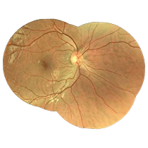
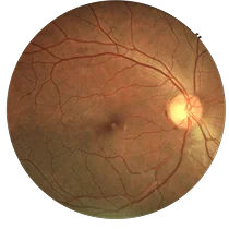
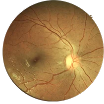
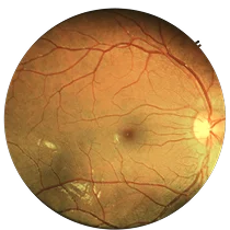
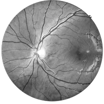
3nethra ultima is a compact, multi-functional digital fundus camera that captures high-resolution 20MP images and supports both non-mydriatic and mydriatic imaging.
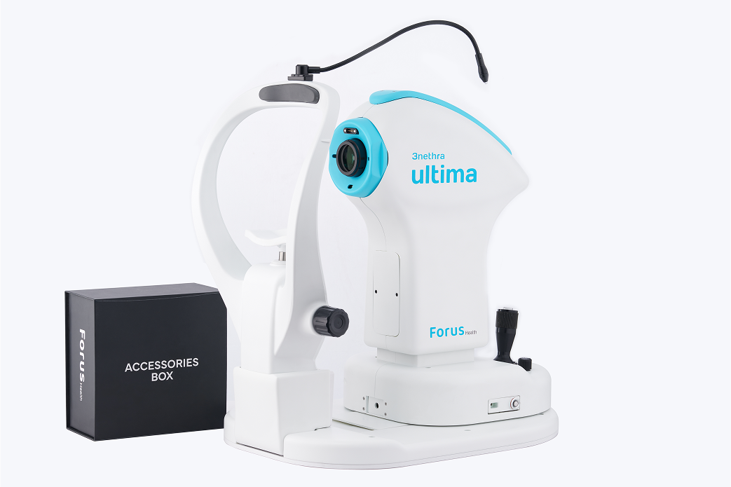
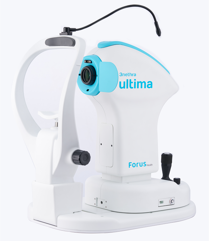
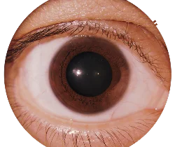
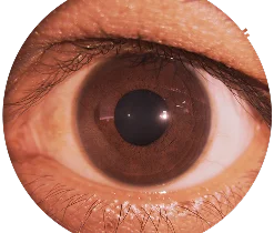
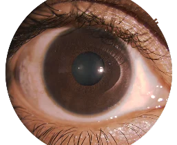
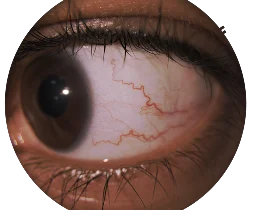
Designed for efficiency and ease of use, it delivers excellent fundus and dry eye imaging with advanced, non-invasive analysis tools. The device automates the detection of glaucoma, diabetic retinopathy, ARMD, cataract, demodex, and pink eye—enhancing clinical workflow and diagnostic accuracy. Additionally, FH-POISE™ enhances AI-driven capabilities for faster, more accurate screenings. With telemedicine integration, 3nethra ultima makes remote eye care more accessible and efficient.
Clinical Highlights
-
Dual Imaging Modes with 20MP Ultra-High-Resolution
Captures exceptionally detailed retinal and anterior segment images in both non-mydriatic and mydriatic modes (with or without dilation), ensuring diagnostic flexibility, patient comfort, and precise visualization to support accurate diagnosis.
-
Wide Field of View with Montage* Capability
Offers a 45° field of view, expandable up to 85° through montage*, enabling comprehensive assessment of the retina and peripheral regions.
-
HDR Imaging for Enhanced Visualization
High Dynamic Range imaging improves detail visibility, especially in the optic disc and peripheral areas.
-
Precise Patient Alignment
Integrated 9-point internal fixation system enhances image consistency and patient focus, improving diagnostic accuracy.
-
Seamless Image Capture
Fast auto-focus and intelligent imaging algorithms reduce operator effort and speed up the imaging process.

-
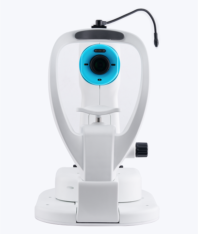
Sleek Monolithic Design
Durable, all-in-one construction enhances ease of use and maintenance in clinical settings.
-
FH-POISE™ Integration
Combines ocular and systemic health insights, offering AI-driven, advanced clinical analysis.
-
FH TeleCare* Compatibility
Enables remote eye screenings and consultations, expanding access through teleophthalmology platforms
-
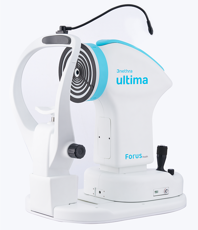
Advanced Non-Contact Dry Eye and Corneal Assessment
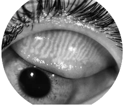
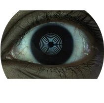
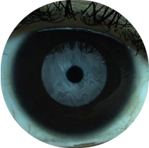
AI-powered tools enable rapid, in-depth and non-invasive dry eye assessment including TBUT, LLI and TMH. Additional assessments include MGD, Fluorescein Imaging, Demodex and Pink Eye.
-
Targeted Pathology Imaging
Supports visualization of specific anterior segment conditions such as Demodex infestation, conjunctivitis (pink eye), and more.
Advantages
Versatile Imaging
Precision and Clarity
Wide Field of View
Compact Clinical Design
Remote Accessibility
Non-Invasive Comfort
Durable Value
Specifications
Image Sensor
- CMOS-based 20 Megapixel
Minimum Pupil Diameter
- ≥ 3 mm
Optical Resolution
- 8-14 microns
Working Distance (Fundus)
- 38 mm
Power Requirement
- USB Type-C, 5V 3A (15W)
Field of View (FOV)
- 45°
Montage*
- 85° (With Internal Fixation)
Refractive Power Compensation
- ± 15 D
Fixation
- External & 9-Point Internal
Focus
- Auto / Manual
Interface
- USB 3.0
Image Format
- PNG, JPEG, DICOM
Observation Light Source
- IR LED
Flash Source
- White LED
FH-POISE™ (Integrated Insights)
- Retinal, Corneal, and Dry Eye Analysis
Dimensions
- 520 mm (H) x 530 mm (L) x 340 mm (W)
Total Weight
- 14 kg with stand
Recommended System Requirements
- Compatible with Windows 11 OS (64-bit) or higher, 16GB RAM or more, Intel i5 processor (9th Gen, 2.4 GHz or higher), 500GB SSD or more, Full HD display (1920 × 1080), USB 3.0 Type-C port with 5V/3A capability.
Forus Health recommends using a CE-marked desktop or laptop. (System specifications are subject to change without notice)
Licensed feature*
